How Choledochal Cysts Are Diagnosed
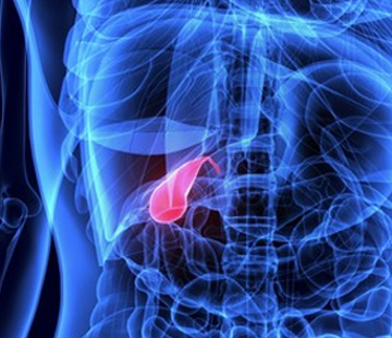
Diagnosing Choledochal Cysts
Being told that you or your child has a choledochal cyst is unsettling, especially when the scans come thick and fast. The reassuring part is that choledochal cyst diagnosis is usually straightforward once the right imaging is done. This page explains each test and what to expect.
How choledochal cysts are found
Before birth, on a routine pregnancy scan
Some cysts are spotted on a routine antenatal ultrasound as a fluid-filled space near the porta hepatis, where the bile ducts leave the liver. StatPearls notes ultrasound can detect these cysts prenatally as a cystic structure in that region.
Nothing usually needs doing before birth. Your team will arrange extra growth scans, plan a normal delivery in most cases, and repeat an ultrasound in the first weeks of life. The cyst must still be confirmed afterwards, because it can turn out to be cystic biliary atresia, a more urgent condition.
After symptoms appear
Many cysts are found later, when pain, jaundice or a lump appears. Cleveland Clinic describes this classic triad of pain, jaundice and a palpable mass, noting most people have only one or two of the three. Our symptoms page has more.
The main imaging tests
Abdominal ultrasound: the first-line test
Ultrasound is nearly always first: sound waves, no radiation, and a good view of a cystic structure connected to the bile duct but separate from the gallbladder. StatPearls reports a sensitivity of 71% to 97%, but adds it alone fails to determine the underlying cause in roughly one in three patients, so it is rarely the end of the story.
MRCP: the modern imaging standard
Magnetic resonance cholangiopancreatography (MRCP) is an MRI technique tuned to make bile and pancreatic fluid glow brightly, mapping the whole duct system. StatPearls calls it the gold standard, with a sensitivity of 90% to 100%, and notes it avoids radiation while still showing the pancreaticobiliary junction. Surgeons use it to plan the operation described on our treatments page.
CT scan
CT uses X-rays and is faster than MRI, useful in emergencies or when someone cannot lie still. StatPearls gives CT cholangiography a sensitivity of about 90%, with the pancreatic duct characterised in 64% of cases. Because of the radiation, it is used sparingly in children.
ERCP: diagnostic, therapeutic, and not risk-free
ERCP passes an endoscope to the duodenum and injects contrast straight into the ducts under X-ray. StatPearls credits it with the highest diagnostic accuracy of any modality, but notes the procedure is now mainly therapeutic, because MRCP and endoscopic ultrasound handle the imaging.
The reason is risk. A systematic review of prospective studies on the NCBI Bookshelf found an overall post-ERCP complication rate of 6.85%: pancreatitis 3.47%, infection 1.44%, bleeding 1.34%, perforation 0.60%, mortality 0.33%. It still earns its place when something needs treating, such as draining an infected duct or opening a choledochocele.
Intraoperative cholangiography
This is a cholangiogram done during surgery, injecting contrast into the biliary tree through the open abdomen or keyhole ports. StatPearls calls it the gold standard for biliary atresia, where failure of contrast to pass confirms the diagnosis.
HIDA scan and ruling out biliary atresia
A HIDA scan uses a radioactive tracer, usually Tc-99m mebrofenin, that the liver excretes into bile. StatPearls reports sensitivity of 100% for type I cysts but only 67% for type IVa, because it detects intrahepatic duct dilation poorly.
Its bigger role in babies is separating biliary atresia from a cyst. If tracer reaches the bowel, biliary atresia is effectively excluded; if not, that is suggestive but not diagnostic, with false-positive and false-negative rates around 10%. This is urgent, because StatPearls reports the Kasai operation works best before 60 days old.
Blood tests and what abnormal results mean
No blood test diagnoses a choledochal cyst, and StatPearls states plainly that laboratory evaluation alone cannot establish the diagnosis. Bloods show how obstructed and inflamed things are.
- Bilirubin: rises when bile cannot drain, causing jaundice and dark urine.
- Alkaline phosphatase (ALP) and GGT: the cholestatic pair, typically raised in obstruction. StatPearls notes GGT has only about 50% to 60% sensitivity and specificity for biliary atresia, so it supports rather than settles a diagnosis.
- ALT and AST: liver cell enzymes, raised when back-pressure or inflammation irritates the liver.
- Amylase and lipase: raised in pancreatitis, a common presentation and a recognised complication.
- Full blood count: a rising white cell count with fever suggests cholangitis, a bile duct infection needing urgent care.
Assigning a Todani type from the images
The cyst is then classified by where the dilation sits. Radiopaedia summarises the five Todani types: type I, dilation of the extrahepatic bile duct, is much the commonest at roughly 80% to 90% of cases; type II is a diverticulum; type III a choledochocele in the duodenal wall; type IV involves intrahepatic and extrahepatic ducts; type V is intrahepatic only, known as Caroli disease.
The type drives the surgical plan. Our guide to the types explains each, with pages on type I cysts and type V, or Caroli disease.
Pancreaticobiliary maljunction and why it matters
In many people with a cyst, the bile and pancreatic ducts join too early, outside the sphincter that normally keeps them apart. StatPearls describes this anomalous pancreaticobiliary junction as the ducts converging 1 to 2 cm before the sphincter of Oddi, in 50% to 80% of cases.
Pancreatic enzymes then reflux into the bile duct and injure its lining, and that chronic irritation is thought to drive the cancer risk. StatPearls puts overall malignancy at about 5%, with a meta-analysis of 2,904 cases reporting 11%, and cholangiocarcinoma appearing at a median age of 49.5 years versus 65 otherwise. Risk was four times higher when a cyst was drained rather than fully excised, which is the argument for complete removal.
What else it could be
Several conditions look similar on a first scan. StatPearls lists a differential including biliary atresia, gallstones in the duct, pancreatic pseudocysts, simple hepatic cysts, duplication cysts and a distended gallbladder. Caroli disease is both a look-alike and, as type V, part of the classification. Ultrasound accuracy for biliary atresia is only around 70% to 80%, which is why a second-line test is standard.
What to expect on the day
Details vary between hospitals, so confirm with your team.
- Ultrasound: a short fast beforehand, often four to six hours in older children, so the gallbladder is not collapsed. No sedation, needles or radiation. Warm gel, a probe under the ribs, 20 to 30 minutes. Your child stays awake and you stay with them.
- MRCP: the British Liver Trust describes this as around 45 minutes, sometimes with a contrast injection into a vein. The scanner is a loud tunnel and images blur with movement, so babies and younger children commonly need sedation or a short general anaesthetic. Older children and adults manage awake.
- CT: quick, roughly 30 minutes, usually with a contrast injection that causes a brief warm flush. It involves radiation, so ask why CT rather than MRCP.
- HIDA scan: StatPearls advises fasting at least four to six hours before the tracer injection, with main imaging about an hour and delayed images sometimes extending to 24 hours. Where biliary atresia is the question, phenobarbital is often given at 5 mg/kg/day for five days beforehand. Babies are usually swaddled rather than sedated.
- ERCP: deep sedation or general anaesthetic, fasting from midnight, several hours in hospital. Ask what pain to report afterwards, since pancreatitis is the main risk.
Bring a comforter and something to distract a hungry child during the fasting window. Our pre-surgery page covers more if surgery is planned.
Questions worth asking after diagnosis
- Which Todani type is it, and how confident are you from the imaging?
- Is there a pancreaticobiliary maljunction, and are the intrahepatic ducts involved?
- Has biliary atresia been fully excluded, and how?
- How many of these operations does this centre do a year?
- What is the timescale, and what symptoms should bring us back sooner?
- What does long-term follow-up involve?
If you are an adult who has just been given this diagnosis, our page on choledochal cysts in adults covers what happens next. Asking for a second opinion at a centre that sees this condition regularly is entirely reasonable. Our find a doctor page helps you locate specialists, our FAQ answers common questions, and our community page connects you with other families.
Please note: this page is general information written for patients and families, not medical advice. Always discuss your own or your child’s diagnosis and results with your medical team.
Sources
- Choledochal Cyst — StatPearls, NCBI Bookshelf
- Choledochal cyst — Radiopaedia
- Endoscopic Retrograde Cholangiopancreatography — StatPearls, NCBI Bookshelf
- Incidence Rates of Post-ERCP Complications — NCBI Bookshelf
- Hepatobiliary Scintigraphy — StatPearls, NCBI Bookshelf
- Biliary Atresia — StatPearls, NCBI Bookshelf
- Choledochal Cyst: Types, Causes and Treatment — Cleveland Clinic
- Choledochal Cysts in Adults — British Liver Trust