Choledochal Cyst Symptoms
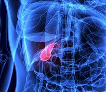
Symptoms
If you or your child has just been told there may be a choledochal cyst, one of the first things you probably want to know is whether the symptoms you have been living with actually make sense. They usually do. A choledochal cyst is a swelling of the bile ducts that has been present since before birth, and almost everything it causes comes back to one thing: bile not draining the way it should.
This page explains choledochal cyst symptoms in plain terms — the classic pattern doctors are taught, why most people do not fit it neatly, how the picture differs between a newborn and a forty-year-old, and which symptoms mean you should be seen the same day.
The classic triad, and why most people do not have all three
Medical textbooks describe a “classic triad” of choledochal cyst symptoms:
- Jaundice — yellowing of the whites of the eyes and then the skin, often alongside pale or clay-coloured stools and unusually dark urine.
- Pain in the upper right abdomen — under the ribs on the right side, where the liver and bile ducts sit. It may be a dull ache, or come in waves.
- A palpable mass — a soft swelling in that same area that a doctor, or sometimes a parent, can actually feel.
Here is the part that surprises many families: the complete triad is seen in only about 20% of cases. So four out of five people diagnosed with a choledochal cyst never have all three signs together. Having only one of them, or something vaguer than any of them, does not make the diagnosis less real. The pattern also shifts with age: among children, roughly 85% present with both an abdominal mass and jaundice, compared with about 25% of adults.
The cyst itself is not always obvious from the outside. Many cysts are found during scans done for another reason entirely, and some cause no symptoms at all until a complication forces the issue. The type of cyst also influences what you feel, because the five types sit in different parts of the biliary tree.
How symptoms differ by age
Before birth: found on a routine ultrasound
A choledochal cyst can sometimes be spotted on a pregnancy scan as a cystic structure near the porta hepatis — the area where the bile ducts leave the liver. If this happens, there are no symptoms to speak of yet; the baby is usually born well and monitored closely. Importantly, a cyst seen before birth cannot be reliably told apart from biliary atresia on prenatal imaging, which is why babies detected this way are assessed promptly after delivery. Our diagnosis page explains the scans involved.
Newborns and babies
In infants under twelve months, the most common picture is jaundice, pale (acholic) stools and vomiting. Parents often describe:
- Jaundice that does not clear up — yellowing that persists beyond two weeks of age is not normal newborn jaundice and always deserves prompt evaluation.
- Stools that turn pale, chalky, putty-coloured or almost white.
- Urine that is dark, sometimes staining the nappy yellow-brown.
- Poor feeding, fussiness during or after feeds, and vomiting.
- Slow weight gain or failure to thrive.
- A firm or enlarged liver, or a swelling on the right side of the tummy.
Pale stools plus dark urine in a jaundiced baby is a combination worth photographing and showing your doctor. It points to bile not reaching the gut, which is exactly what an obstructing cyst does.
Older children
Children past infancy often present less dramatically. Intermittent tummy pain, nausea, episodes of jaundice that come and go, and sometimes a first attack of pancreatitis are typical. Because the pain can settle for weeks at a time, it is easy to attribute it to constipation, a stomach bug or anxiety about school. Around two-thirds of choledochal cysts are diagnosed before the age of ten.
Adults
About a quarter of choledochal cysts are found in adulthood, and that proportion has been rising as imaging becomes more common. Adults most often report right upper quadrant abdominal pain rather than jaundice, and the symptoms tend to be non-specific:
- Recurrent upper abdominal pain, sometimes worse after fatty meals.
- Bloating, a swollen tummy, nausea and reduced appetite.
- Episodes of jaundice or pale stools that resolve on their own.
- Repeated attacks of pancreatitis with no obvious cause.
- Bouts of cholangitis — bile duct infection — with fever and chills.
- Itching, unexplained weight loss, or simply feeling unwell in cycles.
Choledochal cysts are rare — roughly 1 in 100,000 to 150,000 live births in Western countries, and far more common in East Asian populations — and they affect females about four times as often as males. Rarity is a large part of why adults wait so long for an answer. We have a dedicated page on choledochal cysts in adults, covering how the picture differs and why misdiagnosis is so common.
When to seek urgent care
Some symptoms are not wait-and-see symptoms. The combination of fever, jaundice and right upper abdominal pain is known as Charcot’s triad and suggests acute cholangitis, an infection of the bile ducts that can become life-threatening quickly. Only about a quarter of people with cholangitis have all three signs, so do not wait for the full set. Go to an emergency department, or contact your surgical team directly, if you or your child has:
- Fever or shaking chills together with abdominal pain or yellowing.
- Jaundice that appears suddenly or deepens over a day or two.
- Severe or unrelenting upper abdominal pain, especially with vomiting.
- Confusion, drowsiness, dizziness or a racing heartbeat alongside any of the above — these suggest the infection is affecting the whole body.
- In a baby: fever, persistent jaundice, pale stools, or refusing feeds.
Tell whoever sees you that a choledochal cyst is known or suspected. It changes how urgently they will investigate. Our complications page covers cholangitis, pancreatitis and stone formation in more depth.
Why the diagnosis is often missed in adults
Adult symptoms overlap almost perfectly with far more common conditions. Recurrent right-sided pain with nausea looks like gallstones, and many adults have their gallbladder removed before anyone questions the bile ducts. Cyclical bloating and abdominal discomfort get labelled as irritable bowel syndrome. Unexplained pancreatitis may be blamed on alcohol or diet. Because the condition affects so few people, it is genuinely not the first thing most clinicians think of, and standard ultrasound does not always show the cyst clearly. Many adults describe years of appointments before an MRCP finally gives them a name for it. If you are looking for a clinician with relevant experience, our find a doctor resource is a starting point.
Symptoms that appear or persist after surgery
Surgical removal of the cyst with reconstruction resolves symptoms for most people, but the biliary system has been permanently rerouted, so some symptoms can return later. Scarring at the join between the bile duct and bowel (an anastomotic stricture) can cause bile to pool, which in turn leads to sludge and stones, repeat episodes of cholangitis, and sometimes pancreatitis. These can appear years or even decades after the operation. There is also a small residual cancer risk even after complete excision, which is why lifelong follow-up is recommended rather than optional.
Recurring fever, jaundice or right-sided pain after surgery should always be reported — it is not something to push through. Our pages on preparing for surgery and life after the operation go through what recovery normally looks like, and the treatments page explains the procedures themselves.
How to describe your symptoms to your doctor
Vague symptoms get taken more seriously when they are recorded precisely. Before your appointment, try to note:
- Pattern and timing — how often episodes happen, how long each lasts, and whether they follow meals.
- Stool and urine colour — take photos. “Pale stools and dark urine” is a specific clinical signal, not a vague complaint.
- Temperature readings — an actual number during an episode is far more useful than “I felt feverish”.
- Skin and eye colour — photograph the whites of the eyes in daylight when yellowing appears.
- Weight — for children, bring the growth chart; for adults, note any unintentional loss.
- Previous scans and blood tests, and whether an MRCP has ever been done. Ask directly: could this be a bile duct problem rather than a gallbladder one?
Hearing how other families described things can help you find the words. You may find our frequently asked questions, the stories shared by other patients, and our community useful for that.
Please note: this page is general information written to help you understand and describe symptoms. It is not a diagnosis and cannot replace assessment by a qualified doctor. Symptoms listed here have many possible causes, and only imaging and clinical examination can confirm a choledochal cyst.
Sources
- Choledochal Cyst — StatPearls, NCBI Bookshelf
- Choledochal Cyst: Types, Causes & Treatment — Cleveland Clinic
- Choledochal Cysts and Other Biliary Malformations — Children’s Hospital of Philadelphia
- Choledochal Cyst — Seattle Children’s Hospital
- Choledochal Cysts in Adults — British Liver Trust
- Cholangitis — StatPearls, NCBI Bookshelf
- Biliary Atresia — StatPearls, NCBI Bookshelf