Choledochal Cysts Type III
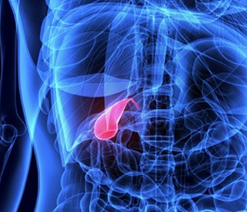
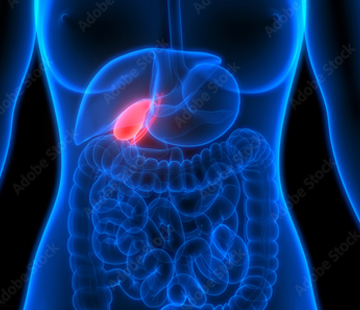

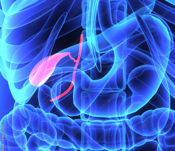
Type III Choledochal cysts, also known as choledochoceles, make up roughly 5% of all bile duct cysts. They are characterized by a protrusion of a focally dilated, intramural segment of the distal common bile duct into the duodenum, the first part of the small intestine.
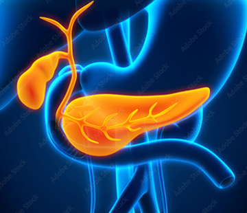
These cysts are typically located within the wall of the duodenum or pancreas. The exact cause of their development remains unknown, but some researchers suggest that they may arise from dysfunction of the sphincter of Oddi, the muscle responsible for controlling the flow of bile and pancreatic juice into the small intestine.
Symptoms of Type III Choledochal cysts can include abdominal pain, nausea, vomiting, and jaundice. Complications may encompass cholangitis, pancreatitis, and biliary obstruction.
Diagnosing Type III Choledochal cysts usually involves imaging studies such as ultrasound, CT scan, or magnetic resonance cholangiopancreatography (MRCP). Endoscopic retrograde cholangiopancreatography (ERCP) may also be employed for diagnosis and treatment.
Treatment options for Type III Choledochal cysts depend on the severity of the condition and the presence of symptoms. Asymptomatic patients may not require treatment in some cases. For symptomatic patients, choledochoceles can be managed with endoscopic sphincterotomy, which involves cutting the muscle of the sphincter of Oddi, surgical excision, or a combination of both approaches.
In summary, Type III Choledochal cysts, or choledochoceles, represent approximately 5% of all bile duct cysts. They are characterized by a protrusion of a focally dilated, intramural segment of the distal common bile duct into the duodenum. Although the precise cause remains unclear, it may stem from dysfunction of the sphincter of Oddi. Early diagnosis and prompt management are crucial for preventing complications and improving patient outcomes.